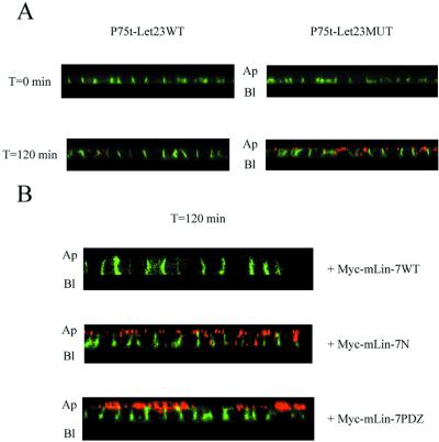
Figure 6.
mLin-7 retained P75t-Let23WT at the basolateral plasma membrane domain in a secondary antibody capture experiment. (A) MDCK cell lines expressing P75t-Let23WT or P75t-Let23MUT were grown on Transwell filters, chilled on ice, and then incubated for 1 h with HEPES-buffered growth medium containing anti-P75 at the basolateral side to bind P75t-Let23 at the cell surface. After washing to remove unbound antibody, the cells were warmed to 37°C in growth medium containing goat anti-mouse IgG-Texas Red conjugate (the capture antibody, shown in red) at the apical side only. At the time indicated to the left, the cells were washed to remove unbound antibody, fixed and permeabilized, stained with goat anti-mouse IgG-FITC conjugate (shown in green) to reveal anti-P75 not already bound to the Texas Red conjugate, and examined by confocal laser-scanning microscopy. The panels show Z-sections that have been contrast enhanced for presentation, and the apical (Ap) and basolateral (Bl) sides are indicated. (B) Similar to experiment described in A, but performed with MDCK cell lines coexpressing P75t-Let23WT with Myc-mLin-7WT, Myc-mLin-7N, or Myc-mLin-7PDZ (see Figure 3 for description of mLin-7 constructs). Only the 120-min time point is shown.

