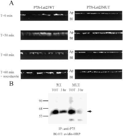
Figure 7.
P75t-Let23WT sorted to the basolateral plasma membrane domain after endocytosis from the apical surface. (A) To examine endocytic sorting, MDCK cell lines expressing P75t-Let23WT or P75t-Let23MUT were grown on Transwell filters, chilled on ice, and then incubated for 1 h with HEPES-buffered growth medium containing anti-P75 at the apical side to bind P75t-Let23 at the cell surface. After washing to remove unbound antibody, the cells were warmed to 37°C in growth medium. At the time indicated to the left, the cells were fixed and permeabilized, stained with goat anti-mouse IgG-FITC conjugate, and examined by confocal laser-scanning microscopy. Nocodazole (20 μg/ml) was added to the incubations and washes for the samples indicated. The panels show Z-sections that have been contrast enhanced for presentation, and the apical (Ap) and basolateral (Bl) sides are labeled. (B) Biotinylation-antibody capture: MDCK cell lines expressing P75t-Let23WT (WT) or P75t-Let23MUT (MUT) were grown on Transwell filters, labeled at the apical side with biotin, and then incubated for 3 h with growth medium containing anti-P75 at the basolateral side to capture biotinylated P75t-Let23 chimera. After washing to remove unbound antibody, the cells were lysed and the lysates subjected to precipitation with rabbit anti-mouse IgG and protein A-Sepharose. The precipitate was separated by 10% SDS-PAGE, transferred to nitrocellulose, and probed for biotinylated proteins with avidin-HRP. Control precipitations were also performed to determine total biotinylated P75t-Let23 (TOT). Relative molecular weight is shown to the left in kilodaltons. An arrow indicates the relevant band.

