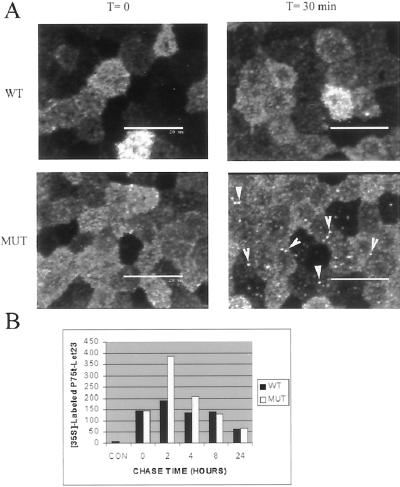
Figure 9.
P75t-Let23MUT was sorted to a degradative pathway after internalization. (A) Endocytosis of chimera proteins from the apical side as described in Figure 7, except that 25 μM chloroquine, a weak base used to counter vesicle acidification, was included throughout the course of the experiment. The initial localization of prebound anti-P75 is shown (T = 0 min). After 30 min at 37°C (T = 30 min), intracellular vesicular staining was evident in P75t-Let23MUT (MUT), but not in P75t-Let23WT (WT). Arrows highlight some vesicles. Bars, 25 μm. (B) Amount of P75t-Let23WT (WT) and P75t-Let23MUT (MUT) remaining over time after pulse-labeling with [35S]methionine was determined by immunoprecipitation with anti-P75 antibody. Chase time in hours is shown on the x-axis, and the amount of labeled protein remaining (determined by densitometry) is shown on the y-axis. Negative control precipitation by using anti-Myc antibodies at T = 0 h is indicated as CON.

