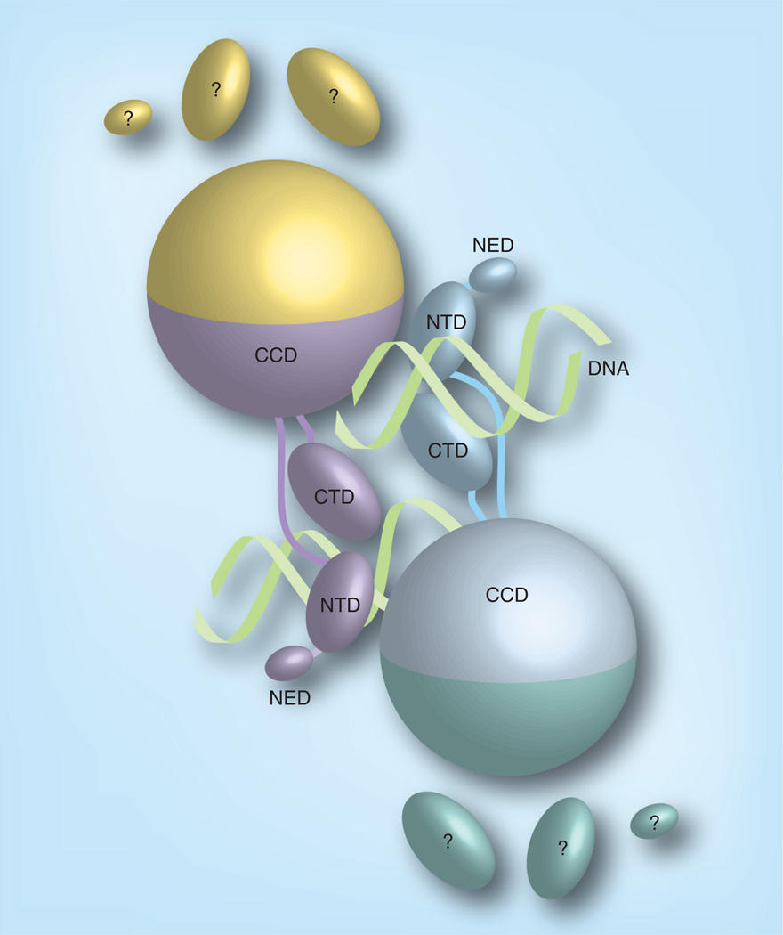
Figure 3. Cartoon representation of the prototype foamy virus intasome structure.
The integrase monomers in the intasome are distinguished by their colors. The CCD, NTD, CTD and NED are distinguished by shape. The pair of viral DNA ends are represented as helices. All the contacts between integrase and viral DNA are with the inner subunits. The CTD, NTD and NED of the outer subunits are disordered.
CCD: Catalytic core domain; CTD: C-terminal domain; NED: N-terminal extension domain; NTD: N-terminal domain.
Adapted with permission from [81].

