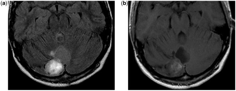
Figure 1.
An 11-year-old boy with a cerebellar pilocytic astrocytoma. (a) Axial T2-weighted FLAIR magnetic resonance (MR) image shows the cerebellar superficial hyperintense nodule and the lack of suppression of the signal in the cystic component. Compare with the CSF signal in the temporal horns. (b) Axial contrast-enhanced T1-weighted MR image shows enhancement of the nodular component and slight enhancement of the cyst wall.

