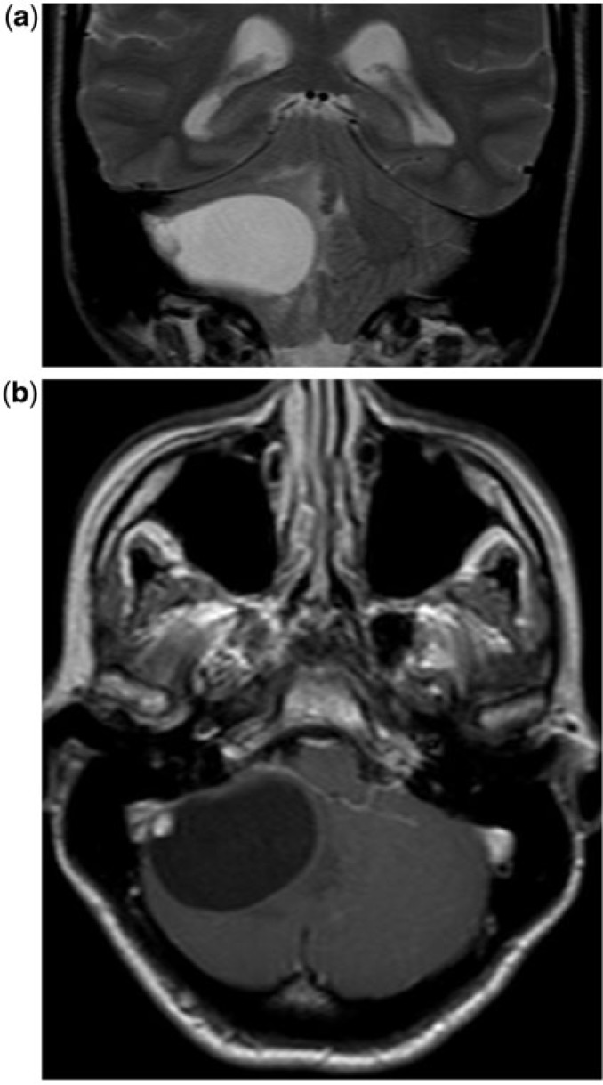
Figure 4.
A 65-year-old woman with a hemangioblastoma. (a) Coronal T2-weighted MR image shows a cyst with mural nodule lesion characterized by a very large cyst and a tiny peripheral nodule, which is hyperintense in T2. The cyst content is slightly hyperintense in T1 compared with CSF. (b) Axial contrast-enhanced T1-weighted MR image show the enhancement of the small nodule that abuts the inferior cerebellum surface and the lack of enhancement of the cyst capsule. The cyst content is slightly hyperintense in T1 compared with CSF.

