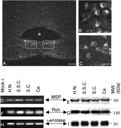
Figure 1.
Ron localization in adult brain. (A–C) Cryostat sections reacted with anti-Ron antibodies and observed with a confocal scanning microscope. Asterisk labels the fourth ventricle. The gray squares in A (bar, 80 μm) encompass the region showed at high magnification in B and C (bar, 33 μm). The antibody stains the hypoglossal motoneurons. Transcripts (D, F, and H) and proteins (E, G, and I) from hypoglossal nucleus (H.N.), somatosensory cortex (S.S.C.), superior colliculus (S.C.), and cerebellum (Ce.). cDNAs were amplified with primers specific for MSP (D), RON (F), and γ-enolase (H) and then loaded onto a 4% agarose gel. Mock r. (first lane) is the result of a PCR performed without template. E, G, and I are Western blots showing the expression, respectively, of MSP, Ron, and γ-enolase proteins in the same animals examined by PCRs. The protein extracts were loaded onto 8% SDS-PAGE gels. The gels were blotted and the filters were decorated with specific antibodies. The molecular weights are indicated (MW kDa). Ron and MSP are expressed in all the extracts examined.

