Figure 3.
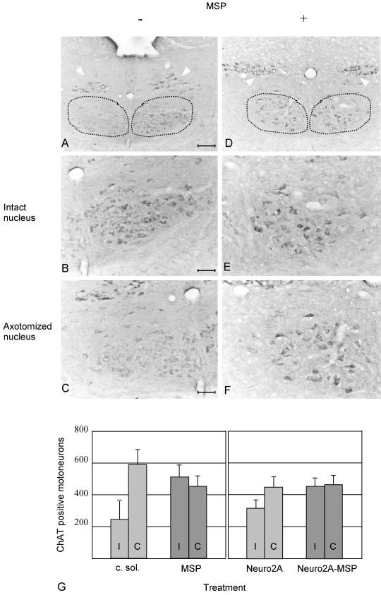
ChAT immunohistochemistry on cryostat sections prepared from axotomized mice treated with conditioned medium of mock-infected Sf9 cells or with MSP. The dotted lines encompass the hypoglossal nuclei; the white arrows point to the dorsal nuclei of the vagal nerve. Mice were sacrificed 1 wk after nerve resection. The hypoglossal nuclei are shown at low (bar, 84 μm, A and D) and high (bar, 33 μm, B, C, E, and F) magnification. In A and D, the nuclei corresponding to the contralateral intact nerves are on the right; on the left are the nuclei of the ipsilateral resected nerves. At high magnification the contralateral nuclei are on the top (B and E) and the ipsilateral on the bottom (C and F). In A–C, mice were treated with conditioned medium of mock-infected Sf9 cells. In D–F, animals were infused with MSP. ChAT activity is lost in axotomized animals treated with conditioned medium of mock-infected Sf9 cells (C) but not in animals treated with MSP (F). The contralateral hypoglossal nuclei (B and E) and the dorsal nuclei of the vagal nerve (arrowheads) are not affected. (G) Diagram showing the number of ChAT-positive hypoglossal motoneurons in treated mice. Both sides ipsilaterally (I) and contralaterally (C) to axotomy were analyzed. On the left side are the data of mice infused with conditioned medium of mock-infected Sf9 cells (c. sol., 7 mice) or with MSP (MSP, 6 mice). On the right are the data of animals transplanted with Neuro2A cells, expressing or not MSP (Neuro2A and Neuro2A-MSP, 4 mice in each group). The differences between ipsilateral and contralateral sides are significant after axotomy and treatment with c. sol. or Neuro2A (p < 0.05), whereas MSP deliver protects the axotomyzed motoneurons from ChAT decline.

