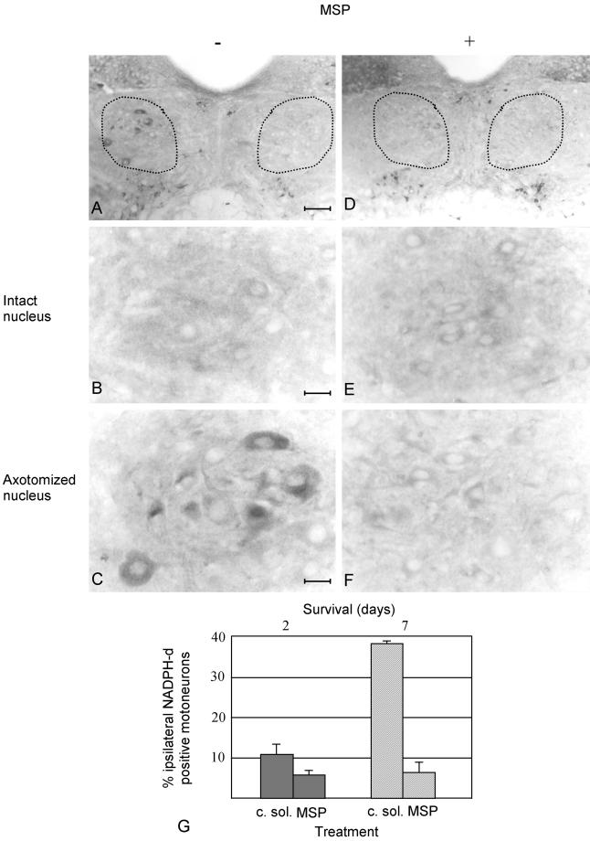
Figure 4.
NADPH-d activity in sections of axotomized hypoglossal nuclei from animals treated with conditioned medium of mock-infected Sf9 cells or with MSP. Mice were sacrificed 2 d or 1 wk after nerve resection. Four mice were analyzed in each group. The dotted lines encompass the hypoglossal nuclei. Panels are presented as in Figure 3. The hypoglossal nuclei are shown at low (bar, 74 μm, A and D) and high (bar, 16 μm, B, C, E, and F) magnification. In A and D, the nuclei corresponding to the contralateral intact nerves are on the right; on the left are the nuclei of the ipsilateral resected nerves. At high magnification the contralateral nuclei are on the top (B and E) and the ipsilateral on the bottom (C and F). In A–C, the animals were treated with conditioned medium; in D–F with MSP. These representative sections are obtained from animals sacrificed 1 wk after axotomy. The contralateral intact sides do not show NADPH-d activity (compare B and E), whereas it is visible in the nucleus ipsilateral to axotomy in animals treated with conditioned medium. This NADPH-d activity is absent when MSP is supplied (F). (G) Diagram of NADPH-d activity 48 h (filled shapes) and 1 wk (dashed shapes) after axotomy, in animals treated either with conditioned medium of mock-infected Sf9 cells (c. sol.) or with MSP. In each animal three to seven brain sections were analyzed. The neuronal cell profile was used as counting unit. NADPH-d-positive motoneurons were counted separately in the nuclei contra- and ipsi-lateral to axotomy and were expressed as percentage of the total number of hypoglossal motoneurons of the corresponding side. The values of the axotomized sides were obtained by subtracting the percentage of the positive motoneurons in the contralateral intact sides to the number of positive motoneurons in the ipsilateral side. The differences in DADPH-d reactivity between axotomyzed nuclei treated with c. sol. and MSP are significant (p < 0.05).

