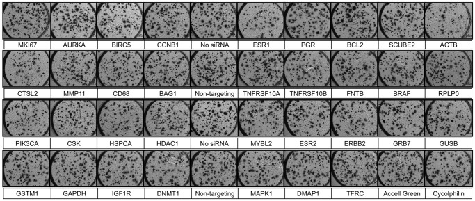
Figure 2. Representative images showing MCF7 colonies growing on the Test Cancer BioChip in presence of individual siRNAs.
Cells were stained with MTT after 15 days on the CBC-1. Live colonies take up the dye and thus appear dark and slightly larger due to the formation of formazan crystals. Each well is 3 mm in diameter.

