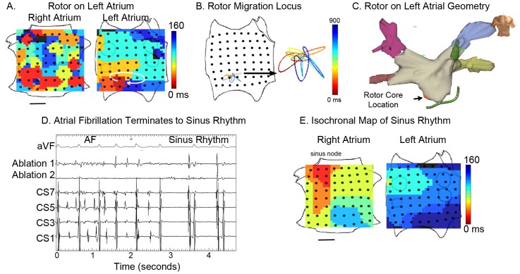
Figure 3. AF termination by ablation of Stable LA rotor.
A. Left atrial rotor during paroxysmal AF visualized using isochrones. B. Migration locus of the rotational center, color-coded over time. C. Ablation lesions at rotor in low left atrium, applied 1 hour after initial recording of the rotor, shown on patient specific geometry. Red lesion is where AF terminated, and 3 other lesions (gray) were also applied. D. Electrode recordings during AF with termination to sinus rhythm by <1 minute ablation at the rotational center (ECG lead aVF, and electrodes at ablation catheter, coronary sinus). E. Isochronal map of sinus rhythm. The patient remains free of AF on implanted cardiac monitor at 9 months. Scale Bar 1 cm.

