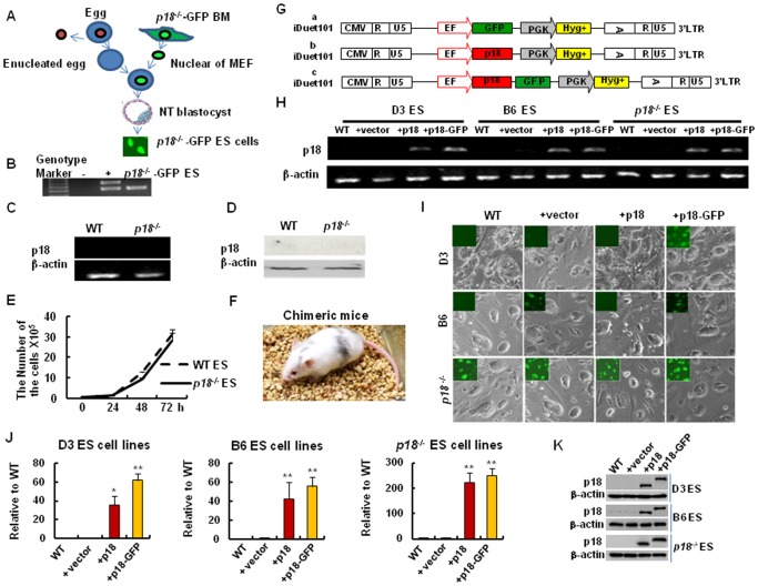
Figure 1. Establishment of “loss-of-function” and “Gain-of-function” models.
(A) Generation of p18−/− ES cells by nuclear transfer. Briefly, the nuclei of p18−/− BM cells were microinjected into enucleated oocytes, and nuclear transfer (NT) embryos developed into blastocysts. These blastocyts were selected for derivation of p18−/− ES cells. (B) The genotype analysis of p18−/− ES cell line. (C) RT-PCR assays to detect mRNA levels of p18 in wild type (WT) and p18−/− ES cells. (D) Protein expression of p18 in WT and p18−/− ES cells detected by western blotting. β-actin was used as a loading control. (E) Growth curves for WT and p18−/− ES cells determined by counting the number of cells present at each time point using trypan blue staining. (F) Chimeric mice were generated by injecting p18−/− ES cells into diploid blastocysts. Reconstituted embryos were then developed in the uteri of foster mothers and chimera pups were obtained 19 days after injection. (G) Schematic representation of the lentiviral vectors used in this study. The vector, iDuet101, contains an EF1 promoter that drives the expression of GFP, p18, or a p18-GFP fusion protein. CMV, cytomegalovirus; R, repeat region in the viral long terminal repeat; U5 regions in the viral long terminal repeat; EF, elongation factor 1α; GFP, green fluorescent protein gene; PGK, mouse phosphoglycerate kinase promoter; Hyg+, hygromycin resistance gene; LTR, long terminal repeat of lentiviral DNA. (H) RT-PCR detection of mRNA levels in mouse ES cells transduced with p18-GFP or p18. Briefly, transduced cells for both groups were selected with hygromycin-B (I), and then infected with the iDuet101-GFP, as well as iDuet101-p18, or p18-GFP, lentiviruses. Top panel (D3 ES), middle panel (B6 ES), and lower panels (p18−/− ES) represent bright field images obtained, as well as fluorescence microscopy images added as inserts. WT: parental ES cells; +vector: iDuet 101-GFP; +p18: iDuet 101-p18; and +p18 - GFP: iDuet 101-p18 - GFP transduced ES cells. (J) Real-time RT-PCR detection of p18 mRNA in mouse ES cells transduced with or without p18 or p18-GFP. Data were analyzed according to the ΔCT method. Values are expressed as the mean ± SD from two independent experiments, and all values were normalized to levels of β-actin. (K) Western blot analysis for p18 expression in three different ES cell lines with or without p18 or p18-GFP overexpression. *, p18-GFP. In B-E, H-K, data represent three independent experiments with similar results.

