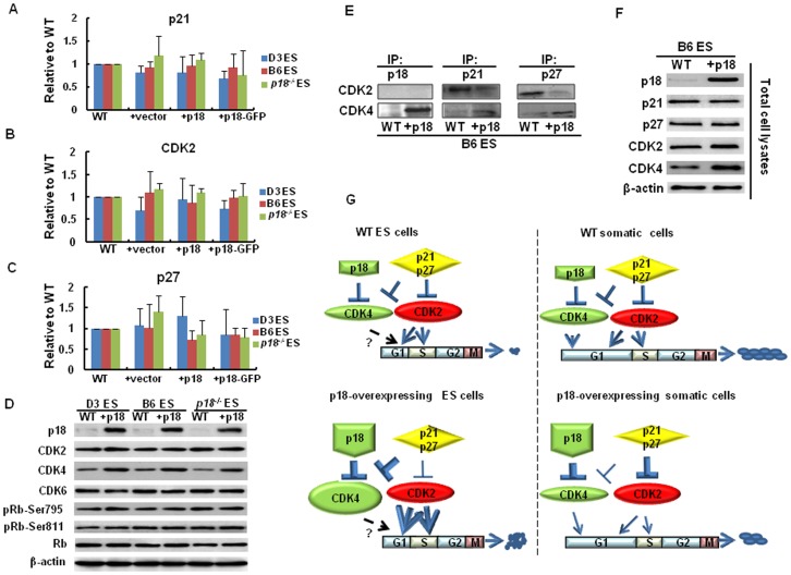
Figure 5. Reassortment of CDKI to CDKs by p18 overexpression in ES cells.
(A, B and C) Real-time RT-PCR analysis of p21, p27, and CDK2 mRNA levels. All values were normalized to β-actin. Values are expressed as the mean ± SD. (D) Western blotting performed to detect protein levels of p18, CDK2, CDK4, CDK6, pRb-Ser795, pRb-Ser811, and total Rb. Detection of β-actin was used as a loading control. (E) IP assays of p18, p21 and p27 were performed using cell lysate (100 µg total protein) from stably transduced p18, or WT ES cells. Immunocomplexes obtained were then immunoblotted with anti-CDK4 and anti-CDK2 antibodies. (F) Total protein extracts were obtained from stably transduced p18, or WT ES cells and immunoblotted with anti-p18, anti-p21, anti-p27, and anti-cdk2 and anti-cdk4 antibodies. β-actin was used as a loading control. (G) A model for a proposed mechanism by which p18 enhances the self-renewal of ES cells, while inhibiting their differentiation potential. Briefly, ectopic expression of p18 promotes overexpression of CDK4, which in turn enhances the association of p21 and p27 with CDK4, and ultimately upregulates CDK2 activities. As a result, the cell cycle is accelerated and the self-renewal is enhanced, whereas the differentiation process is slowed. In A to F, data represent three independent experiments with similar results.

