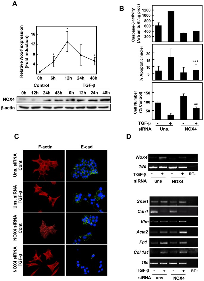
Figure 6. NOX4 is required for apoptosisbut not necessary for the EMT process, induced by TGF-β in hepatocytes.
(A) TGF-β induces NOX4 expression in hepatocytes. NOX4 transript and protein levels were determined by Real time-PCR (upper panel) and western blot (lower panel), respectively, at the indicated times of treatment with 2 ng/ml TGF-β. In B–D, immortalized hepatocytes were transfected with either an unsilencing siRNA (uns siRNA) or a specific siRNA for NOX4 (NOX4 siRNA), serum-depleted for 4 hours and, finally, treated or not with 2 ng/ml TGF-β. (B) Cell death analysis: activation of caspase-3 activity at 16 hours, where the peak of maximal activation was found (upper graph); percentage of cells with apoptotic nuclei at 48 h upon nuclear staining with DAPI (middle graph); loss in cell viability at 48 h (lower graph). Data represent the mean ± SEM of three independent experiments. Student's t test calculated comparing TGF-β-treated cells between the two conditions with the different siRNAs (**p<0.01; ***p<0.001). (C) Fluorescence microscopy staining for F-actin and E-cadherin at 48 hours. (D) Representative RT-PCR of the indicated genes after 3 h of treatment to observe the effects on Nox4 expression (upper panel) and 48 h of treatment to analyze the effects on EMT-related genes (bottom panel). 18S was used as loading control.

