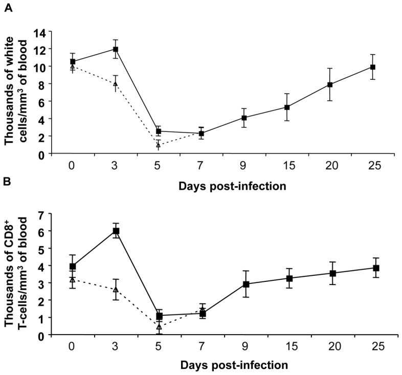
Figure 7. Surviving pigs show a rapid recovery from the ASFV-provoked leukopenia and a dramatic expansion of CD8+ T-cells.

(A) Total nucleated-cell counting using whole blood samples taken at different days after ASFV challenge. (B) Number of CD8+ T-cells found at different times post-infection from these same blood samples after PBMC separation and CD8+ surface specific staining using the specific anti-CD8 monoclonal antibody. Values represented in both panels correspond to the average number of cell per mm3 of pig blood. Standard deviation shown corresponds to those found within the pCMV-control group (dashed line) and those found for the three surviving pigs, pre-immunized pCMV-UbsHAPQ (solid line). No significant differences were observed in the cell counts (nor in the total, nor the specific CD8+ T-cell counts), between non-surviving pigs, independent of the DNA plasmid used.
