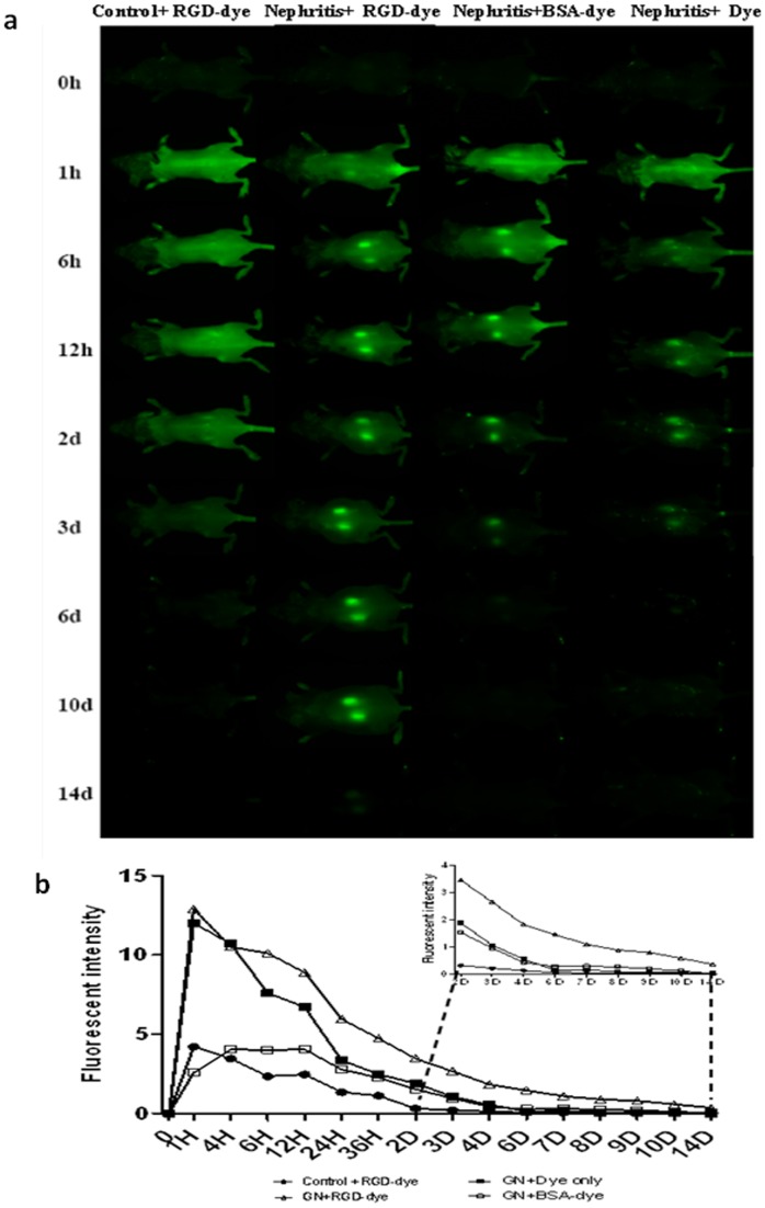
Figure 3. Dynamic in vivo fluorescence imaging using the Pearl® Impulse system.
Repeated in vivo fluorescence imaging was performed using the Pearl® Impulse system over a period of 14 days after 800CW-RGD dye injection. (a) Dynamic renal imaging at successive time points. From left to right: healthy-control + RGD Dye, nephritis + RGD Dye, nephritis + BSA-conjugated dye, and nephritis + Dye only. (b) Average fluorescent signal intensity at each time point for each group, with the data from days D2–D14 shown in the inset (N = 4–5 for each group at each time point, P<0.05 for nephritis + RGD Dye group vs. other group, two-tailed t test). Note: “nephritis” refers to mice that have been challenged with anti-GBM serum.

