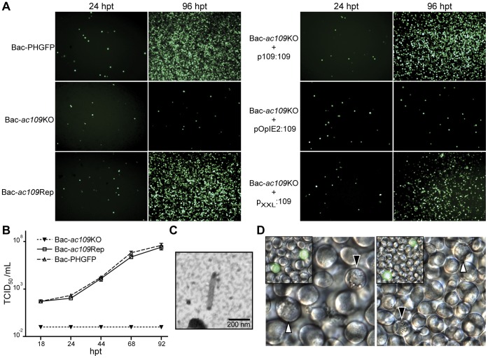
Figure 1. Characterization of Bac-ac109KO and complementation assays.
(A) Fluorescence microscopy of Sf-9 cells transfected with DNA of Bac-PHGFP, Bac-ac109KO or Bac-ac109Rep bacmids (left panels) or cotransfected with Bac-ac109KO bacmid and one of the following plasmids: TOPO-p109-109, pIB109 and pXXL109 (right panels). eGFP fluorescence was monitored at 24 and 96 h post-transfection (hpt). Plasmid TOPO-p109-109 carried ac109 driven by its own promoter (p109∶109), plasmid pIB109 harbored ac109 under constitutive pOpIE2 promoter regulation (pOpIE2∶109) and plasmid pXXL109 carried ac109 driven by a modified version of the very late polyhedrin promoter regulation (pXXL:109). (B) Production of infectious budded viruses (BVs). Sf-9 cells were transfected with Bac-ac109KO, Bac-PHGFP or Bac-ac109Rep bacmids, and at the indicated time points the supernatants were harvested, clarified and tittered by the end point dilution method. Titers were calculated as TCID50 per milliliter. Each sample was performed in triplicate. (C) Electronic microscopy of an ac109KO BV. Sf-9 cells were transfected with Bac-ac109KO bacmid, and 4 days post-transfection culture supernatant was harvested, clarified and concentrated by ultracentrifugation through a 25% w/v sucrose cushion. The pellet was adsorbed to copper grids, negatively stained and analyzed by transmission electronic microscopy. (D) Light (main figure) and fluorescence (left inner figure) microscopy of cells transfected with Bac-ac109KO bacmid. At 96 hpt, cells showing eGFP fluorescence were observed for polyhedra formation. White arrows indicate fluorescent cells without polyhedra and black arrows indicate fluorescent cells with polyhedra.

