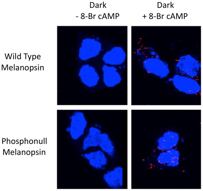
Figure 2. Proximity-dependent ligation assay.

Melanopsin-transfected HEK cells were fixed with 4% PFA for 30 min. with or without pretreatment with 200 µM 8-Br cAMP for 30 minutes before fixation. Melanopsin phosphorylation was assayed with the PLA as described in Materials and Methods. The red fluorescence puncta indicates that the antibody bound to melanopsin’s intracellular C-terminal domain is within 40 nm of the phospho-serine antibody when bound to phosphorylation sites in the intracellular loops. Cells visualized by confocal microscopy. Blue staining indicates DAPI staining of cell nuclei. Images represent Z-stacks of images taken through entire cell.
