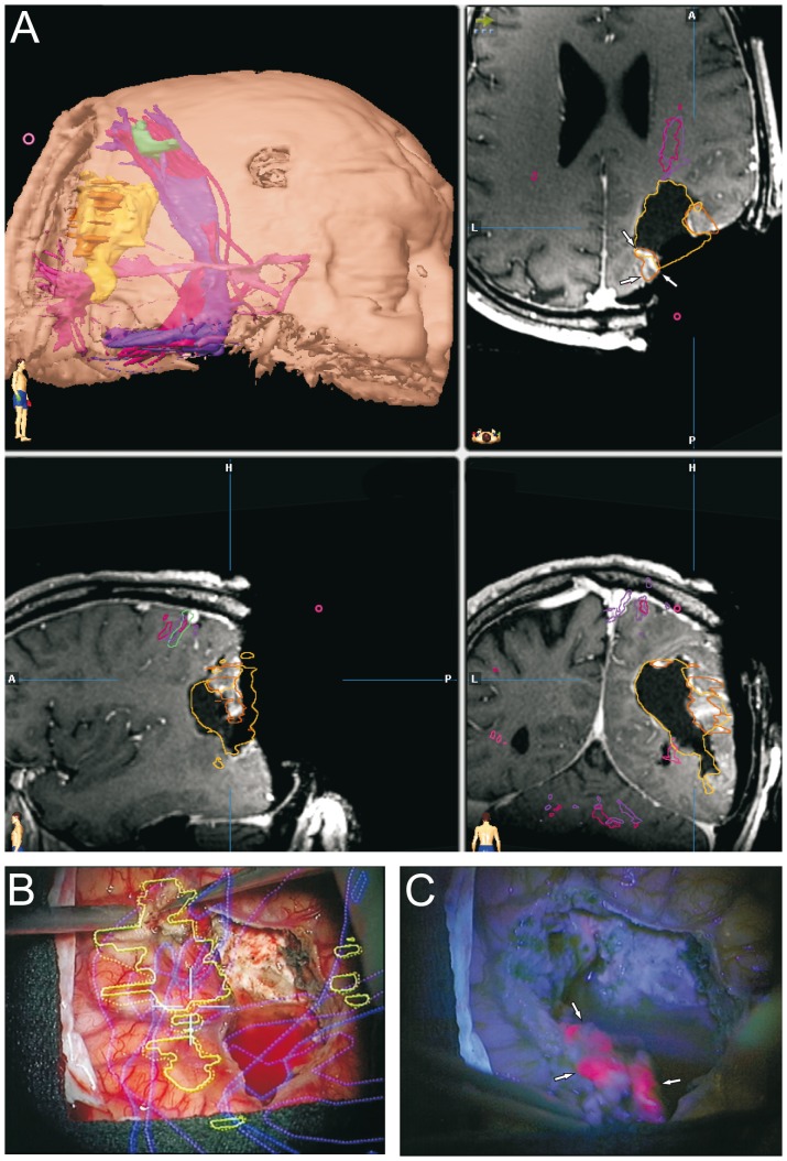
Figure 3. Dual intraoperative visualization approach and anatomical view.
On the basis of a typical case of a tumor in the vicinity of an eloquent area, we demonstrate that the risk of missing tumor remnants covered by non-pathological tissue is eliminated through an iMRI control. A, The first iMRI scan carried out following the disappearance of the 5-ALA signal depicted a residual contrast enhancing area (marked by arrows). B, Tumor resection was resumed following re-segmentation and update of the neuronavigation. C, During resection of the intervening layer of non-pathological tissue, the 5-ALA signal reappeared (marked by arrows) and corresponded to the re-segmented contrast enhancing area.

