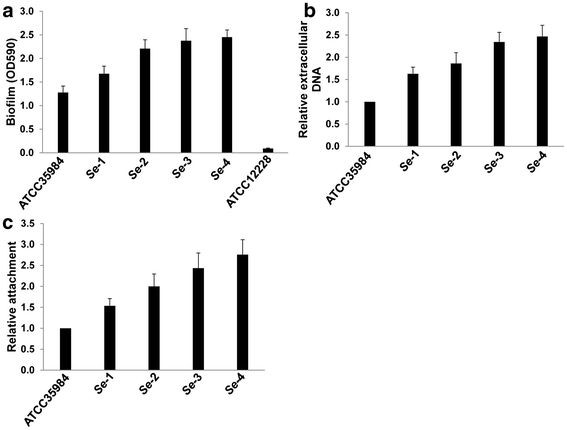
Figure 2.

S. epidermidisisolates associated with catheter infection display more biofilm formation, extracellular DNA release and initial attachment than laboratory strain.(a) Cultures were grown in microtitre plates for 24 h at 37°C, and biofilm biomass was quantified using a crystal violet assay. (b) Cultures were grown for 24 h in minimal medium supplemented with 0.05 mM PI, whereupon PI absorbance (OD480) and cell density (OD600) were measured and relative amounts of extracellular DNA per OD600 unit were calculated. (c) Initial attachment of S. epidermidis strains in static chambers was measured as described in Methods. Error bars represent the S.E.M. for three independent experiments.
