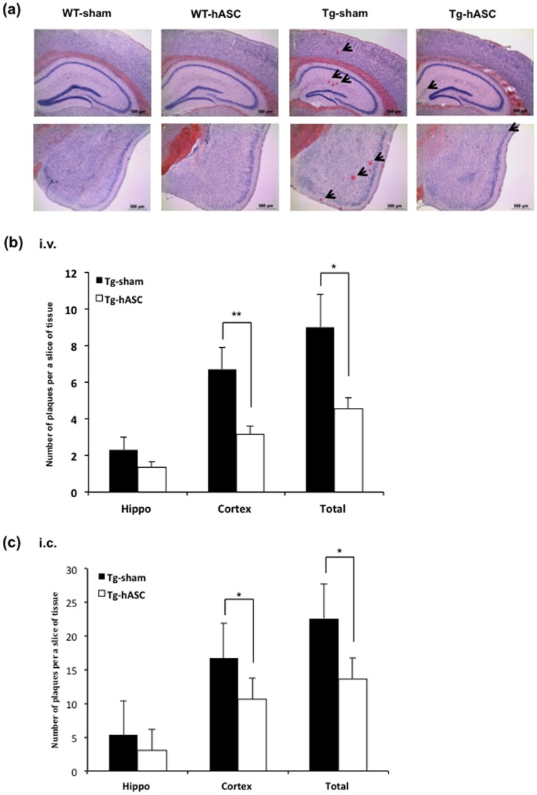
Figure 3. Intravenous and intracerebral injection of hASC reduced the number of amyloid plaques in Tg2576 mouse brains.
(a) Congo red staining for the detection of amyloid plaques was carried out in the hippocampus of each group 4 months after (i.v.) injection. (b) 4 months after the 13th (i.v.) injection, the number of plaques was counted in the hippocampal region of the Tg-hASC and the Tg-sham group. (c) At 4 months after hASC (i.c.) injection, the number of plaques was counted in the hippocampal region of Tg-hASC and Tg-sham groups. All data are represented as mean ± SEM (n = 9∼15 per group). Asterisk *, P<0.05, **, P<0.01 by one-way ANOVA.

