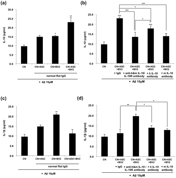
Figure 6. Increased IL-10 might be contributed by hASCs and the secreted level of IL-1β might be modulated by secreted IL-10 from hASCs.
(a) Primary mouse neurons were grown in coated 24-well culture dishes to near confluence 80% in neurobasal media containing B27 for 7 days. They were then added to 10 µM of oligomeric Aβ42 peptides and co-cultured with hASCs and/or BV2 cells. Blocking of IL-10 and IL-10 receptor interaction was performed for 48 h. A neutralizing IL-10 or IL-10 receptor antibody (5 µg/ml, respectively) was used in the indicated groups and IL-10 or IL-1β ELISA was performed. (a–b) The concentration of IL-10 in hASC/BV2 co-culture system was measured with ELISA. (c–d) The concentration of IL-1β in hASC/BV2 co-culture system was measured with ELISA. Data represent mean ± SEM of three independent experiments (n = 30). Asterisk *, P<0.05, **, P<0.01, ***, P<0.001; by One-Way ANOVA: Tukey’s HSD Post Hoc test.

