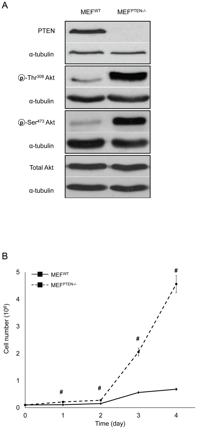
Figure 1. Characterization of MEFPTEN−/−.
(A) Western blot analyses of PTEN, Akt, p-Ser473Akt and p-Thr308Akt Immunoblotting of PTEN, Akt, p-Ser473Akt and p-Thr308Akt was carried out on whole cell lysates of MEFWT and MEFPTEN−/−. The loss of PTEN in MEFPTEN−/− resulted in hyperactivation of Akt on both phosphorylation sites without affecting the expression of total Akt. The respective loading controls of α-tubulin were included. (B) Rate of cell proliferation This was measured by the trypan-blue exclusion assay. The MEFPTEN−/− has a significantly higher rate of proliferation compared to MEFWT from days 1–4 of culture. # p<0.005 for n = 3.

