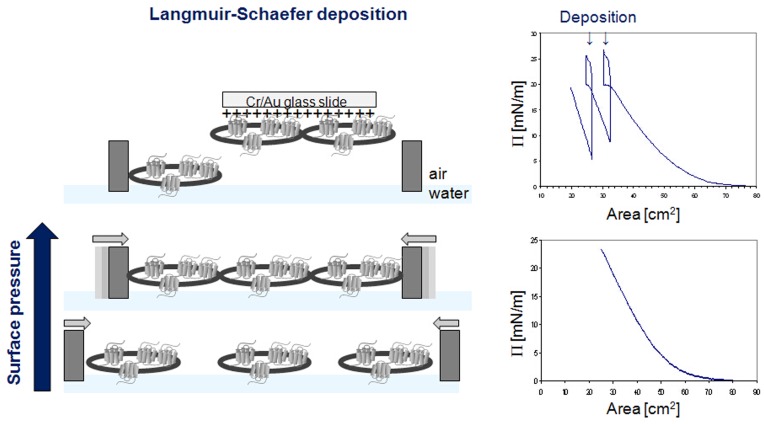
Figure 4. Langmuir-Schaefer deposition of human cells.
Cell droplets were spread over the water surface in the Langmuir trough, and the trough barriers were moved towards each other to compress the cells; the surface pressure diagrams are shown on the right hand side. At a surface pressure Π of 20 mN/m - just before pressure saturation when cells are present in a uniform layer - Cr/Au coated glass slides carrying a positive charge were brought in to contact with the cells on the water surface and removed again, transferring a portion of the cell layer onto the slide.

