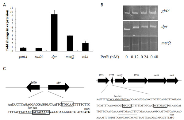
Figure 3.
Identification of PerR regulon inS. suis. (A) Relative expression levels of genes dpr, metQ, relA, pmtA and sodA in strain ΔperR compared to its parental strain SC-19. Relative abundance of the transcripts was determined by real-time RT-PCR from the total RNAs derived from strains ΔperR and SC-19 in mid-log phase. gapdh was used as the internal control. (B) Different concentration of PerR proteins binds to dpr and metQIN promoters (500 bp and 300 bp respectively), gidA promoter (300 bp) was used as the negative control. (C) The structure of the dpr and metQIN promoters. -10 and −35 regions of the promoters are shown by the boxes. The start codon is labeled by blod fonts. The predicted PerR-box is underlined.

