
Figure 9.
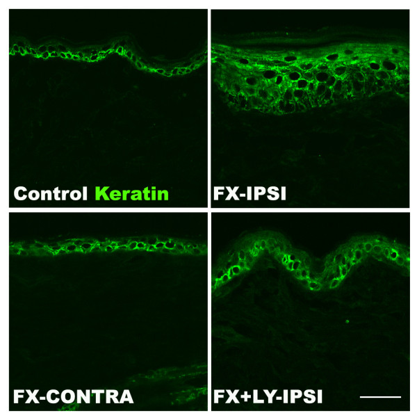
Immunostaining for keratin in the hindpaw skin four weeks post-fracture. Panels exhibit confocal images from the hindpaw skin of a normal control rat, the ipsilateral hindpaw skin of a fracture rat (FX-IPSI), the contralateral hindpaw skin of a fracture rat (FX-CONTRA), and the ipsilateral hindpaw skin of a fracture rat treated with an NK1 receptor antagonist (LY303870 20 mg/kg, i.p. daily for 8 days, FX + LY-IPSI), showing staining for keratin (green). Scale bar = 25 μm. There was an increase in epidermal thickness, indicating increased keratinocyte proliferation, at 4 weeks post-fracture in the fracture paw (FX-IPSI), but not in the contralateral paw (FX-CONTRA), and this increase was blocked in fractured rats treated with LY303870 (FX + LY-IPSI).