
Figure 7. Effects of β-AR stimulation on content of free thiols of RyRs.
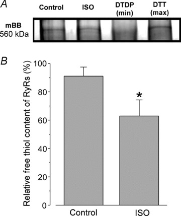
A, mBB fluorescence intensity signals of RyRs measured under control conditions and after 15 min of ISO (0.1 mm) treatment. For both groups, myocytes were electrically stimulated at 0.75 Hz. The maximal and the minimal free thiol content were determined after treatment of cells with DTT (5 mm) and DTDP (0.5 mm), respectively. B, relative free thiol content of RyRs from control and ISO-treated samples were obtained by normalizing mBB fluorescence to RyR amount determined using Coomassie Blue staining of the gel run in parallel. *P < 0.05 vs. control.