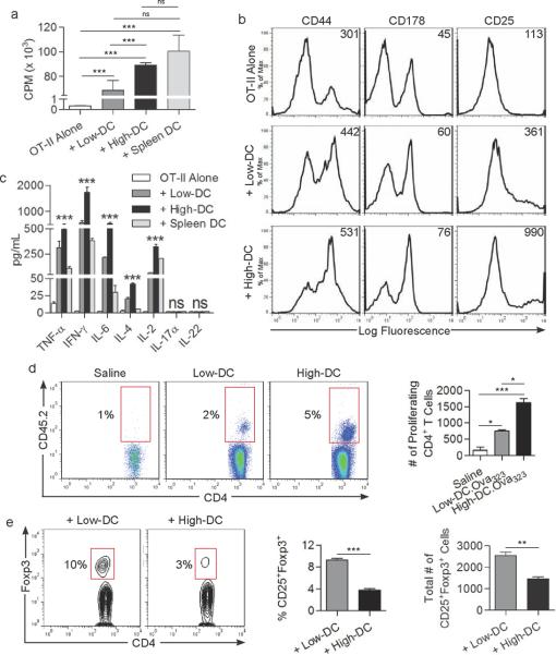
Figure 3. High-DC potently activate of CD4+ T cells whereas Low-DC generate Tregs.
(a) CD4+OT-II cellular proliferation, (b) surface phenotype, (c) and cytokine production was measured after co-culture with High- or Low-DC loaded with Ova323–339 peptide or culture with peptide-pulsed spleen DC. (d) CD4+OT-II T cell proliferation was also measured in vivo in the draining popliteal lymph node after footpad immunization with saline, or antigen-loaded High- or Low-DC. The fraction and total number of proliferating CD4+ T cells is shown. (e) After five days of DC co-culture with allogeneic CD4+ T cells, CD4+CD25+ cells were gated and analyzed for co-expression of Foxp3. The fraction and total number of CD25+Foxp3+ cells is shown. Experiments were performed in triplicate and repeated three times (*P<0.05; **P<0.01; ***P<0.001).

