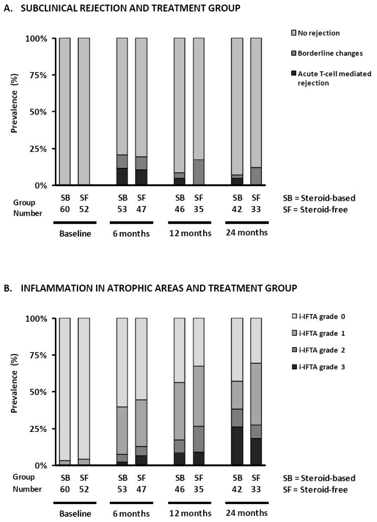
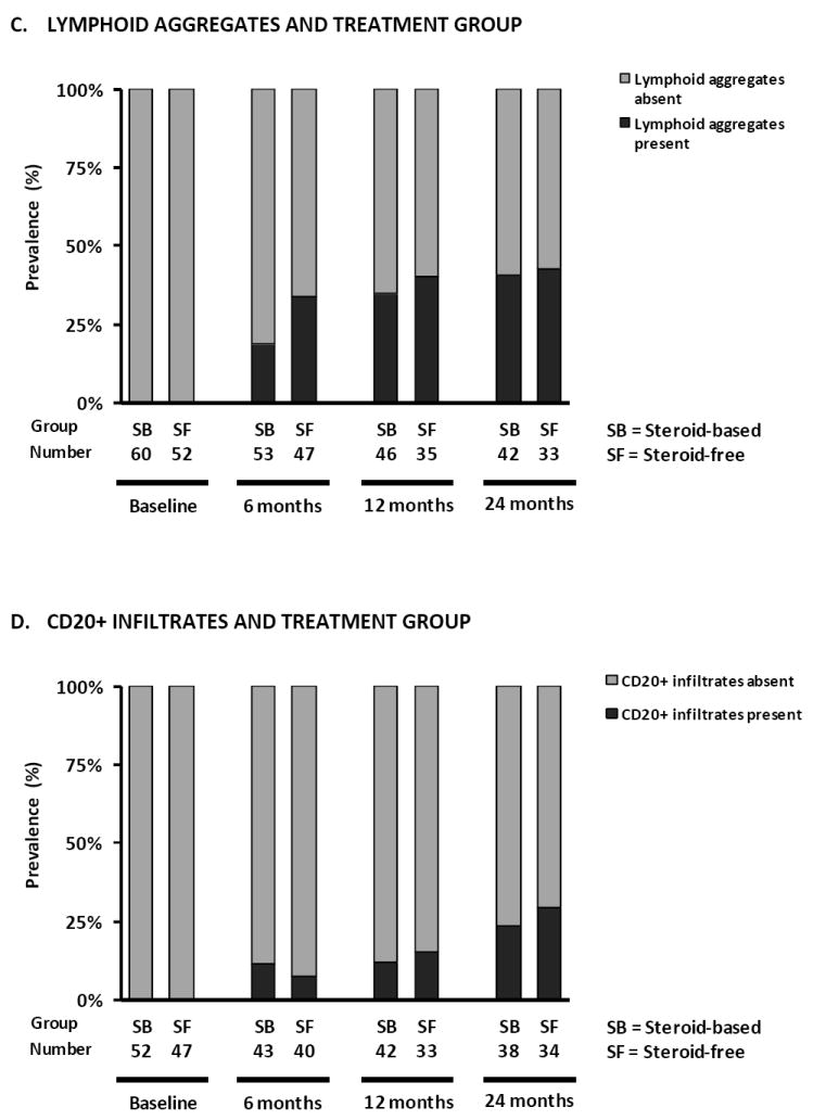
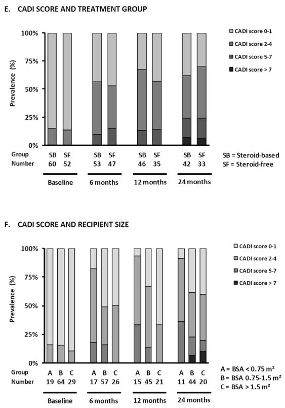
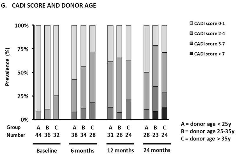
Figure 3. Prevalence of different histological lesions at each time point after transplantation.


Prevalence of subclinical rejection (A), inflammation in atrophic areas (B), lymphoid aggregates (C) and CD20+ infiltrates (D) according to treatment arm in protocol biopsies. (E) Prevalence of different degrees of CADI score according to treatment arm, (F) recipient size and (G) donor age in protocol biopsies. There were no statistically significant differences between the treatment arms in terms of the different histological lesions in GEE analysis (Supplemental Table 3). Smaller recipient size (p<0.0001) and older donor age (p<0.05) were independently associated with higher degrees of CADI score in post-transplantation biopsies in GEE analysis.
