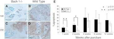
Fig. 9.
Detection of apoptosis cells. TUNEL-stained sections at 1 and 2 weeks after puncture (a–d). Apoptosis cells were stained brown and non-apoptosis cells were stained blue. The number of apoptosis cells increased after puncture in wild-type mice more than in Bach 1−/− mice. Scale bar = 40 μm. The apoptosis rate of Bach 1−/− mice was significantly lower than that of wild-type mice at 1 week (p < 0.01) and 2 weeks (p < 0.05) (e). No significant difference was observed from 4 to 12 weeks after puncture between Bach 1−/− mice and wild-type mice

