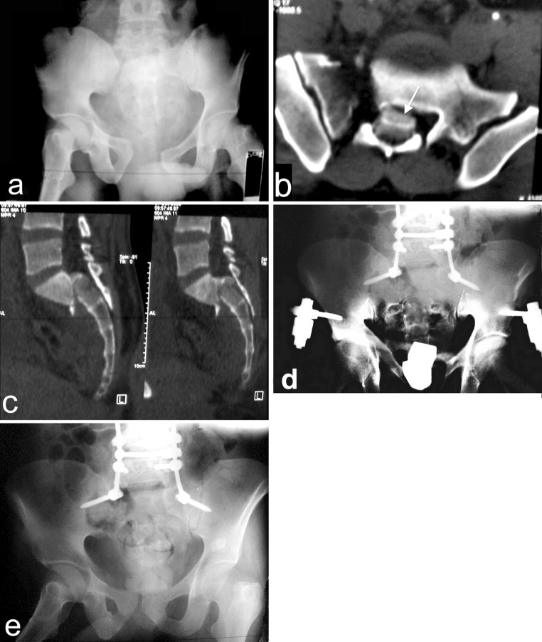
Fig. 6.
a The AP radiograph of 25 years old male patient, he sustained a highly unstable pelvic injury with complete CES (Gibbons type IV). b An axial CT showing Denis type III sacral fracture with sacral canal blocking (white arrow). c The sagittal CT showing type II Roy-Camille spinopelvic dissociation. d The AP radiograph after 1 month from direct posterior decompression and lumbopelvic fixation. e The AP radiograph after 11 months from surgery showing good fusion. The CES regressed after 20 months to Gibbons type II

