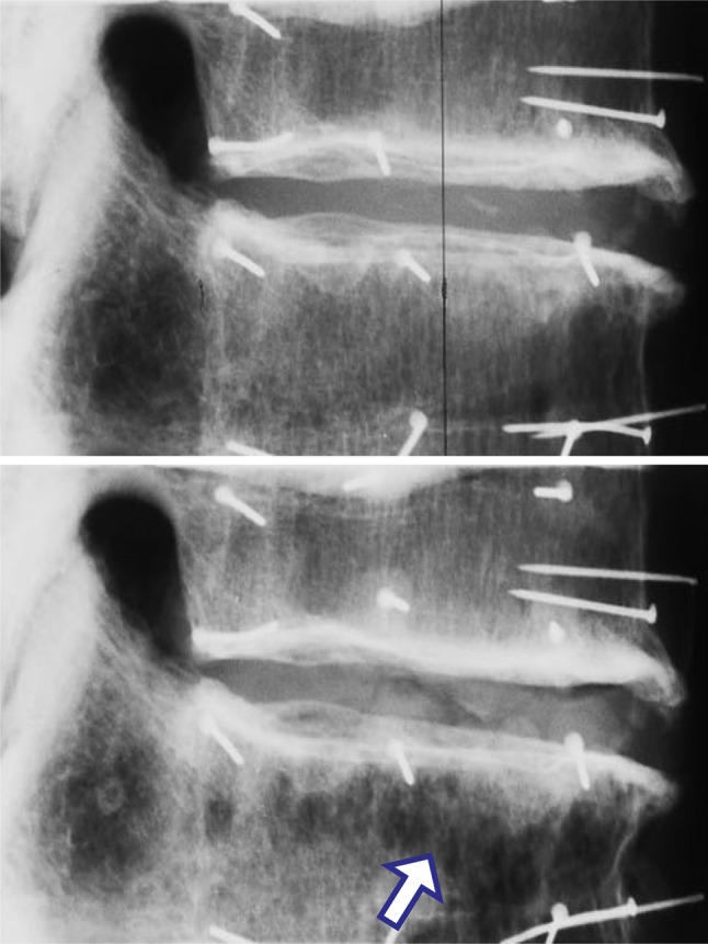
Fig. 5.

Lateral radiographs of a two-vertebra specimen, before (upper) and after (lower) minor compressive damage (male 66 years, T10-11, anterior on the right). In the lower radiograph, note the disturbed anterior trabeculae (arrow). Damage reduced the height of this specimen by 2.29 mm. Pins were inserted into the vertebrae for the attachment of reflective markers
