Figure 2.
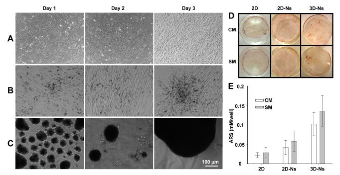
Morphological changes and mineralization of VSMCs. Cells were incubated in control media (CM), or in stimulating media (SM) supplemented with 50 μg/ml ascorbic acid (AA), 7.5 mM β-glycerophosphate (β-GP), and 10 nM l-α-lyso-phosphatidylcholine (LPC) for 4 days. Morphology of VSMCs in 2D (A), 2D-Ns (B), or 3D-Ns (C) SM cultures. Cells were washed with PBS and observed under light microscopy. Images of cells cultured in CM are shown in Supplemental Figure II. (D) Cells cultured in CM or SM were stained with Alizarin Red-S (ARS) to detect the calcium mineral layer and images captured by a digital camera after the removal of unbound ARS dye by washing with water. (E) Mineral layers stained with ARS in CM or SM were quantified at 405 nm (results are mean ± SD, n = 4).

