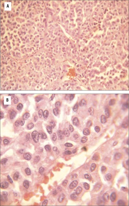
Figure 4. Eosin and hematoxylin staining of adrenal adenoma (40X magnification) showing small round adrenal tumor cells with prominent nuclei and nucleoli, bands of fibrous septae separating the tumor cells, without evidence of vascular invasion; Figure 4b: High magnification (100X) showing blue round adrenal cells with increased nucleo-cytoplasmic ratio and prominent nucleoli and no evidence of vascular invasion.

