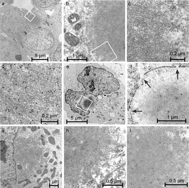
Figure 7.
Electron micrographs of 293 Tet-Off cells containing HDQ83 aggregates. After HDQ83 expression for 3–5 d, cells were fixed as described in MATERIALS AND METHODS and viewed by electron microscopy. (a–c) Different magnifications of a cell containing a typical perinuclear inclusion body. At higher magnifications, HDQ83 fibrils with a diameter of ∼10 nm can be observed. Immunogold labeling of cells with the HD1 antibody confirmed the identity of the HDQ83 fibrils (d). Immunogold labeling of cells also reveals that the proteins ubiquitin (h) and 14–3-3 (i) are present in the interior of inclusion bodies. In cells with perinuclear inclusion bodies, ultrastructural changes such as large indentations of the nuclear envelope (e), disruption of the nuclear membrane (f), and concentration of mitochondria in the vicinity of inclusion bodies (g) are observed. In f, alterations of the nuclear envelope are indicated by arrows.

