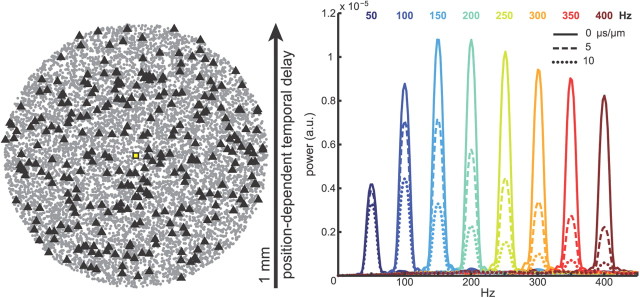
Figure 5.
Effect of spatial synchrony on oscillatory potentials. Left, Locations of CA1 pyramidal cell somata within a 1-mm-diameter disk. The triangles show the location of cells spiking within a 5 ms interval (3% of the population). The temporal offsets of the periodic probability density function that modulates spike timing are shifted in a position-dependent manner along one dimension within the cell body layer, similar to the case in which activity propagates along one direction in CA1 (Lubenov and Siapas, 2009). Right, Average power spectra of Ve in stratum pyramidale over 25 trials with pyramidal neurons undergoing rhythmic firing, as in Figure 2, with varying levels of spatial synchrony. Color indicates the frequency of the firing rhythm, and line type indicates the time delay per unit distance of the oscillating spike probability function. Solid lines, 0 μs/μm (no delays); dashed lines, 5 μs/μm; dotted lines, 10 μs/μm.

