Figure 4.
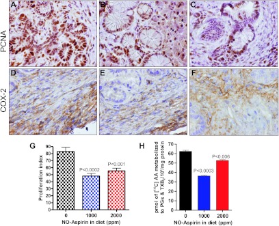
Effect of NO-aspirin on cell proliferation (A–C) and COX-2 (D–F) in pancreatic tumors. Immunohistochemical analysis was performed with paraffin-embedded and microsectioned pancreatic tissues as described in Materials and Methods. Immunohistochemical analysis of PCNA (A–C) and COX-2 (D–F) expression in PanIN lesions and in ductal cells. A significantly decreased expression of PCNA and COX-2 was seen with low-dose NO-aspirin treatment. (G) Significant differences in PIs were observed between the groups. (H) Effects of NO-aspirin on COX activity in the pancreas from mice as assessed with the radio-HPLC method. Values are means ± SEM, N = 6 per treatment group. A significant (P < .006 and 0.0003) inhibition of AA metabolites (PGs and TXB2) was observed in the NO-aspirin-treated group.

