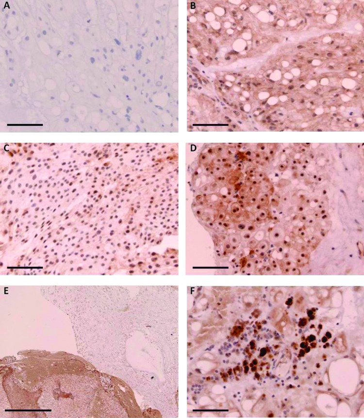
Figure 6.
Immunostaining for FHIT protein in four different classic clival chordomas. (A) Secondary antibody-only control; scale bar, 50 µm. (B) Moderate staining in a region of physaliferous cells, 22-year-old female; scale bar, 50 µm. (C) Minimal cytoplasmic staining and moderate nucleolar staining in a tumor from a 39-year-old female; scale bar, 50 µm. (D) Intense staining of nucleoli and variable cytoplasmic staining in a tumor from a 78-year-old male; scale bar, 50 µm. (E) Regions of moderate cytoplasmic staining juxtaposed with regions of no cytoplasmic staining in a recurrent tumor from a 44-year-old male; scale bar, 0.5 mm. (F) Intense cytoplasmic and nuclear staining in tumor-associated macrophages; scale bar, 50 µm.

