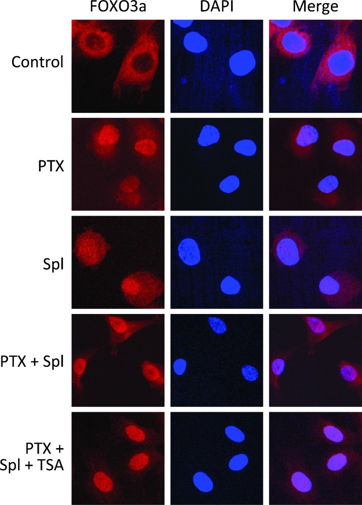
Figure 5.
Confocal microscopy analysis of the effect of paclitaxel and tubulin deacetylase inhibitors on the localization of FOXO3a in endothelial cells. HUVECs were exposed to paclitaxel (PTX, 10 nM), splitomicin (Spl, 1.2 mM), and TSA (1 µM) for 4 hours, processed, and immunostained for FOXO3a as detailed in Materials and Methods. Scale bar, 10 µm.

