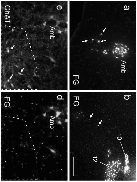
Fig. 2. The ChAT-ir neurons of the mRVLM are neither motor neurons nor parasympathetic neurons.
a Fluorogold (FG) neurons within the compact subdivision of the ambiguus nucleus (Amb; left side of the brain) and surrounding reticular formation (loose formation of Amb.; arrows; approximate coronal level 6.6 mm caudal to bregma). FG natural fluorescence was visualized with an excitation filter of 365 nm and emission filter of 420 nm. These FG-labeled neurons are for the most part motor neurons that innervate the esophagus and airways and may include a few parasympathetic preganglionic neurons. b FG-labeled neurons within the dorsal medulla (obex level; left side of medulla oblongata). Hypoglossal nucleus (12) and dorsal motor nucleus of the vagus (10) are strongly labeled. Putative parasympathetic preganglionic neurons located within the intermediate reticular formation are indicated by arrows. c,d higher-power photographs of the mRVLM region (approximate bregma level −7.0 mm). c ChAT immunoreactivity visualized with Cy3 (excitation filter 545 nm, emission filter 605 nm). d FG illustrating the absence of FG in the mRVLM-ChAT-ir neurons (region outlined by the dotted line). ChAT-ir neurons belonging to the loose formation of the nucleus ambiguus can be seen dorsolaterally to the mRVLM. These neurons exit the CNS and contain FG as expected. Scale bar = 200 μm (a,b), 100 μm (c,d).

