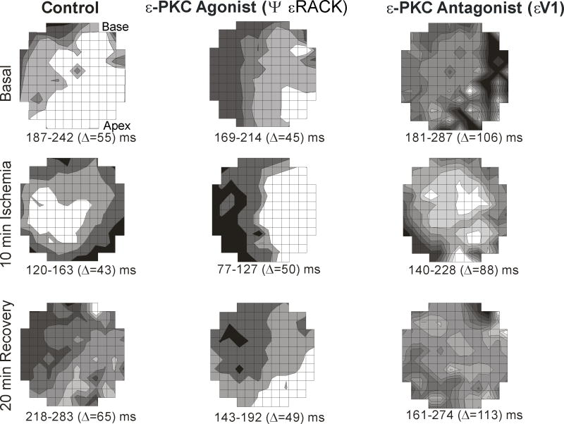
Figure 3. Maps illustrating spatial distribution of action potentials recorded from guinea pig hearts.
The maps were obtained at 10 ms isochrones of action potential duration at 90% (APD90) during basal conditions, 10 min ischemia and 20 min reperfusion. During basal conditions (prior to ischemia), the control untreated hearts and εPKC agonist ψεRACK treated hearts, APD90 had less dispersion of 55 ms and 45 ms, respectively compared with εPKC antagonist εV1 treatment (106 ms). During ischemia, the εPKC antagonist εV1 treatment resulted in the worst spatial heterogeneity of the shortening of APD90 with the highest dispersion (88 ms) compared to control (43 ms) and εPKC agonist ψεRACK (50 ms). At the end of the 20 min reperfusion, εPKC agonist ψεRACK) treatment resulted in full recovery of APD90 dispersion (49 ms) similar to the dispersion prior to ischemia under basal conditions (45 ms). However, in the εPKC antagonist εV1 treated hearts, APD90 dispersion was the worst (113 ms) compared to untreated control (65 ms) and to εPKC agonist εV1 (49 ms).

