Figure 7.
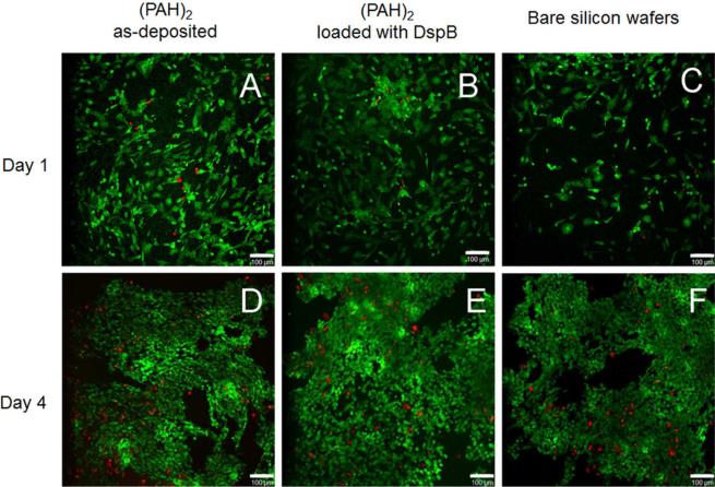
Confocal laser microscopy images of HFOB 1.19 cell attachment and proliferation at as-synthesized (PAH)2 coatings (A and D), DspB-loaded (PAH)2 films (B and E), as well as at the surface of bare silicon wafers (C and F) after 1 day (A, B, C) and 4 days (D, E, F). Representative live/dead images indicate much higher number of live cells (green) as compared to dead cells (red). The scale bar is 100 μm.

