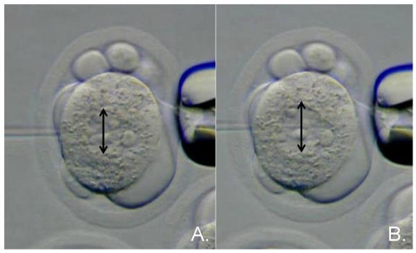
Figure 6.

DNA microinjection into the male pronucleus of the one-cell mouse embryo. (A.) Injection needle moving into the male pronucleus of the one-cell embryo. (B.) Expansion of the male pronucleus upon delivery of DNA under hydrostatic pressure. The black arrows show that the pronucleus diameter increases in size by approximately 50%.
