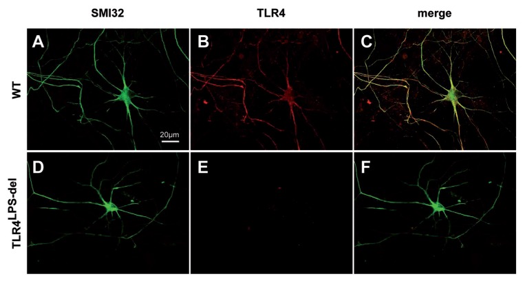
Figure 1.
TLR4 expression on motor neurons from WT or TLR4LPS-del mice. Motor neuron/glia cocultures from WT (A, B) or TLR4LPS-del (D, E) mouse embryos underwent immunocyto-chemistry for SMI32 (A, D, green) and TLR4 (B, E, red). Images were acquired by a confocal microscope. WT motor neurons showed a widespread TLR4 distribution colocalizing with SMI32-positive dendrites and axons (C, merge). TLR4LPS-del motor neurons gave only a very weak, aspecific red fluorescence (E). Scale bar, 20 μm.

