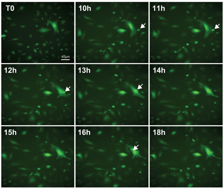
Figure 3.
Live imaging analysis of microglial activation induced by LPS. Purified microglial cultures were incubated with a GFP-lentiviral construct for 72 h. The success of the infection was first confirmed by optical microscopy, and the cultures under the different experimental conditions were then analyzed by a time-lapse recording. Pictures at different time points from the recording of a LPS-treated (1 μg/mL) culture are reported, showing cell debris phagocytosis by an activated microglial cell (white arrows). Scale bar, 40 μm.

