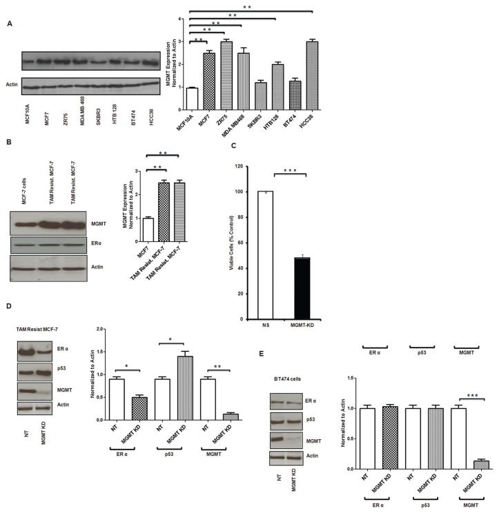
Figure 2.
MGMT protein expression in breast cancer cells. MGMT expression was determined by Western blot analysis. (A) MGMT expression in normal epithelial breast cells was compared with several breast cancer cell lines. Breast cancer cell lines showed higher expression of MGMT compared with normal breast epithelial cells (MCF10A). (B) Tamoxifen-resistant MCF7 breast cancer cells show significantly increased MGMT expression compared with MCF7 parental cells (B). (C) Effect of MGMT inhibition by specific siRNA was determined on tamoxifen-resistant MCF7 breast cancer cell growth by ATP assay. Tamoxifen-resistant MCF7 cells were transfected with NT (nontarget) and MGMT siRNA (MGMT KD); and ATP assays were performed after 72-h treatment. MGMT-silenced cells showed more than 50% decreased luminescence when compared with NT cells. (D, E) Inhibition of MGMT by siRNA led to inhibition of ERα expression and increased p53 expression in wt p53 cells (tamoxifen-resistant MCF7 cells) and there was no change in the mild ERα positive, mutated p53, low MGMT expressing BT474 cells.

