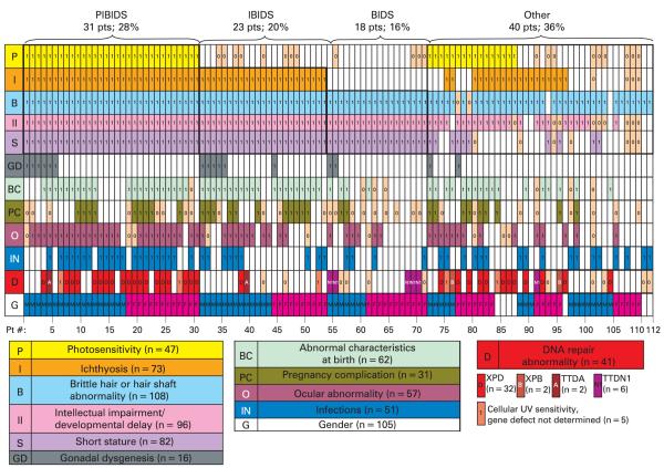
Figure 4.
A clinical array of features reported in the literature on 112 trichothiodystrophy patients. Each column of rectangles represents clinical features of one reported patient. Presence or absence of each feature is indicated in each rectangle of a column. Abnormal clinical features reported are indicated by “1” in a coloured rectangle. Normal reported features are indicated by “0” in a tan rectangle. Unreported features are blank. The rows represent P (yellow)—photosensitivity (n = 47 cases); I (orange)—ichthyosis (n = 73 cases); B (powder blue)—brittle hair or hair shaft abnormality (n = 108); II (pink) —intellectual impairment (n = 96 cases); GD (grey) —gonadal dysgenesis (n = 16 cases); BC (light green) —abnormal birth characteristics (n = 62 cases); PC (dark green) —pregnancy complications (n = 31 pregnancies); O (maroon)—ocular abnormality (n = 57 cases); IN (royal blue) —infections (n = 51 patients); D (red)—DNA repair abnormality (n = 41); D in red rectangle—XPD (n = 32 cases); B in striped red rectangle—XPB (n = 2 cases); A in striped red rectangle—TTDA (n = 2); N in striped pink rectangle—TTDN1 (n = 6 cases); I in red rectangle—cellular ultraviolet (UV) hypersensitivity, gene not determined (n = 5); G—gender (n = 105 patients); blue rectangles—males (n = 54 cases); pink rectangles—females (n = 51 cases). Patients whose clinical features fulfil the criteria for PIBIDS (28%), IBIDS (20%), BIDS (16%) and those that do not (OTHER) (36%), are grouped by bold outline (decreased fertility is ignored in this grouping due to inability to assess in children.)

