
Figure 3. Labeling of non-diabetic and type 2 diabetic induced pluripotent stem cell-derived cardiomyocytes (N-iPSC- and DM-iPSC-CMs, respectively) using a lentiviral vector.
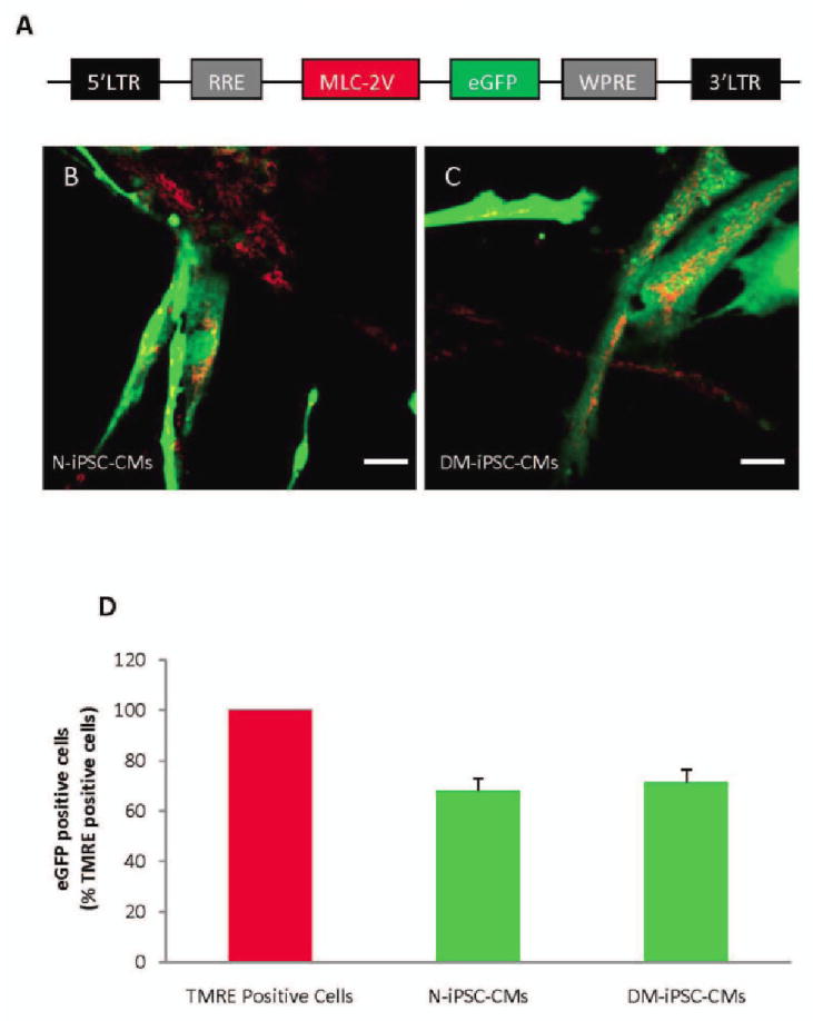
Schematic representation of lentiviral vector pHR(+)C.mlc-2V.egfp.r(−)w(+) used for labeling cardiomyocytes (A). To determine total cell number, cells were loaded with mitochondrial marker tetramethylrhodamine ethyl ester (TMRE, red); myosin light chain-2v (MLC-2v)- enhanced green fluorescent protein (eGFP)-positive cells were counted by detecting green fluorescence, giving the number of cardiomyocytes derived from N-iPSCs and DM-iPSCs (B, C, respectively). Summarized data from six separate differentiations, exhibiting a high percentage of induced pluripotent stem cell-derived cardiomyocytes (D). Scale bar = 40 um.