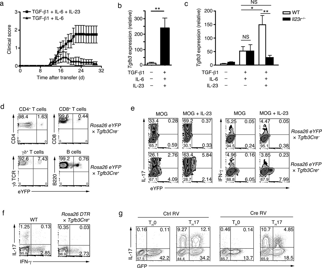
Figure 1. The induction of TGF-β3 in TH17 cells.
(a) Mean clinical scores (disease incidence) in wild-type recipients 30 days after transfer of naïve CD4+ T cells (5×10⁶ cells) from 2D2 transgenic mice that were differentiated in vitro with TGF-β1-IL-6-IL-23 or TGF-β1-IL-6. (b) Quantitative RT-PCR analysis of Tgfb3 mRNA in naïve CD4+ T cells differentiated for four day in vitro with TGF-β1-IL-6-IL-23 or no cytokines. (c) Quantitative RT-PCR analysis of Tgfb3 mRNA in naïve CD4+ T cells from wild-type (WT) or Il23r−/− mice differentiated in vitro with TGF-β1-IL-6-IL-23 or no cytokines. (d) Flow cytometry analysis of TGF-β3-YFP expression in CD4+ T cells, CD8+ T cells, γδ T cells and B cells from the lymph nodes and spleen of TGF-β3crexRosa26yfp mice. (e) Intracellular cytokine staining showing co-expression of TGF-β3-YFP with either IL-17 or IFN-γ in naïve CD4+ T cells from MOG35–55+CFA immunized TGF-β3crexRosa26yfp mice stimulated with PMA+ionomycin four days post in vitro culture with or without IL-23. (f) Intracellular cytokine staining showing IL-17 and IFN-γ expression in PMA+ionomycin ex-vivo stimulated splenocytes from MOG35–55-immunized TGF-β3Cre+xRosaDTR and TGF-β3Cre-xRosaDTR littermate control (WT) mice treated with diptheria toxin on day 4 post-immunization. (g) IL-17 expression in naïve TGF-β3fl/fl CD4+ T cells cultured in vitro with IL-1β-IL-6-IL-23 (TH17) or no cytokines (TH0) then retrovirally transduced with Cre-GFP to delete TGF-β3. All data are a representative of more than three independent experiments with similar results. Statistical significance of *p<0.05, **p<0.01, or ***p<0.001 is indicated for the RT-PCR data. Error bars indicate mean ± s.d.

