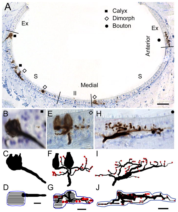
Figure 2.
Lagena receptor epithelium and afferent type. A) Cross section of the lagena along the dorsal-ventral axis of the epithelium, with striola (S), extrastriola (Ex), and type II band (II) regions indicated. Calyx (squares), dimorph (open diamonds), and bouton (circles) afferents filled with BDA neural tracer are shown. B – J) Photomicrographs and anatomical reconstructions of representative calyx (B–D), dimorph (E–G), and bouton (H–J) afferents. Below each photo image lies an anatomical reconstruction drawn in the same plane to scale (calyx terminals = closed contours; bouton terminals = red circles). The second reconstruction represents an apical view of the entire innervation pattern, with contours drawn for the calyx terminals at 1μm focal planes (black lines). Innervation area measurements were obtained from planimeter contours (blue) drawn around the terminal profile. Scale bar = 100μm in A; 5μm in B – J.

